
Major Breakthrough by Chinese Researchers in Asymmetric Protocell Division
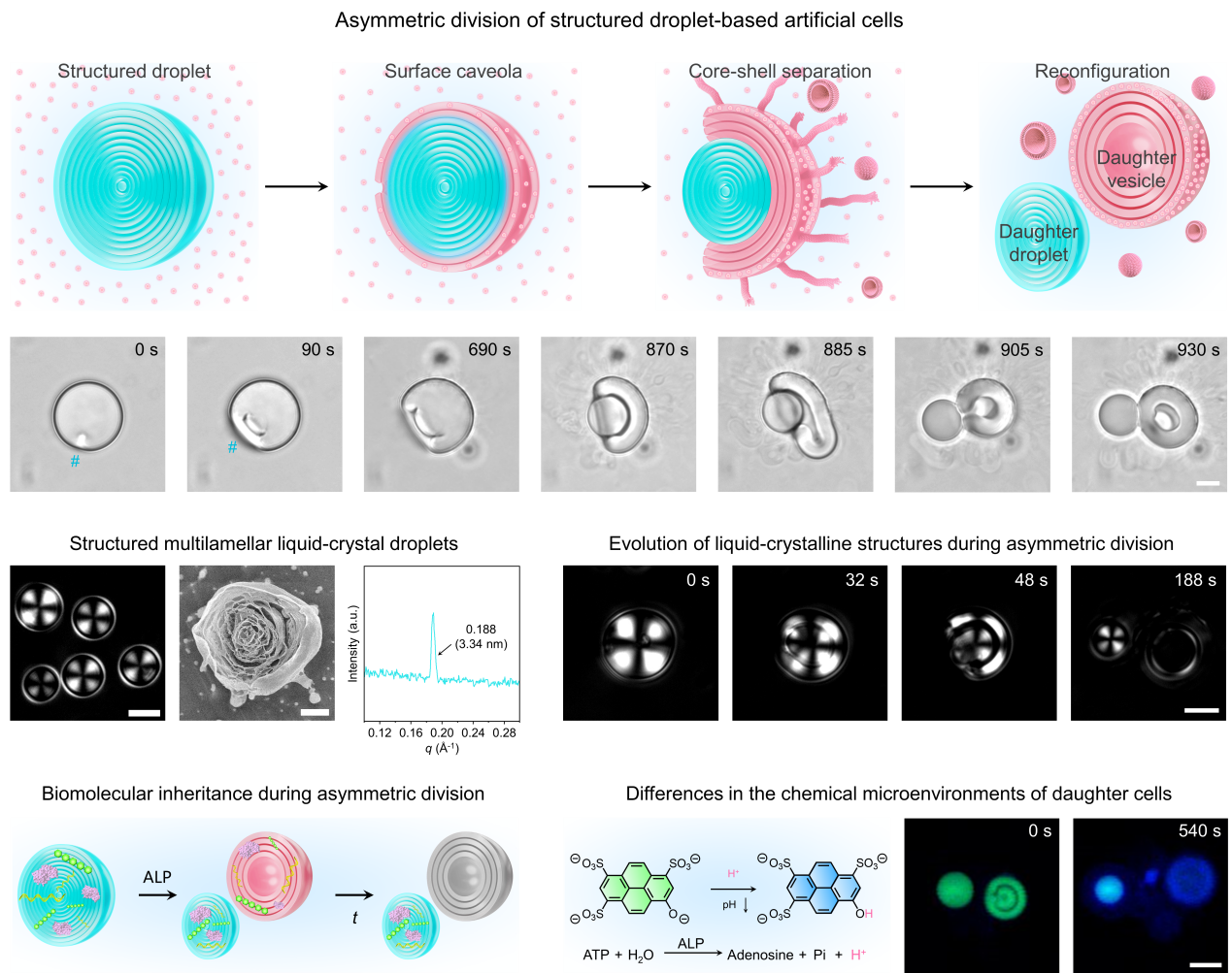
Figure 1. Asymmetric division of structured droplet-based artificial cells
Under the support of the National Natural Science Foundation of China (Grant Nos. T2425001 and 52221006), a collaborative team led by Yan Qiao and Shu Wang from the Institute of Chemistry, Chinese Academy of Sciences, Yiyang Lin from Beijing University of Chemical Technology, and Stephen Mann from the University of Bristol has made important progress in the study of asymmetric division in artificial cells. The research article, entitled “Asymmetric splitting in dividing lipid–nucleotide multilamellar droplets”, was published in Nature on May 13, 2026. Article link: www.nature.com/articles/s41586-026-10489-5.
Controlled division of artificial cells remains a key challenge in mimicking cellular behaviors. To date, researchers have induced vesicle division by modulating membrane tension, membrane fluidity, and osmotic pressure through approaches such as protein machinery, external shear forces, light stimulation, ionic strength regulation, and thermal gradients. Symmetric division in emulsion and coacervate droplet systems has also been achieved through mechanisms including thermal gradients, dissipative self-assembly, wetting energy, and chemical reactions. In contrast, asymmetric cell division is a fundamental process in living systems that underpins cell differentiation, development, and functional diversification. However, owing to the complex spatiotemporal regulation and structural reorganization involved, reproducing this process in artificial cell systems has remained highly challenging.
To address this challenge, the research team developed an innovative strategy for asymmetric division in artificial cells based on transient chemical heterogeneity and interfacial energy gradients, establishing a structured droplet-based artificial cell model with autonomous asymmetric division capability (Figure 1). Under the catalysis of alkaline phosphatase, a localized caveola first forms on the droplet surface. As the enzymatic reaction proceeds, the caveola gradually propagates circumferentially across the droplet surface, while a distinct shell–core interface emerges within the droplet. Once the opening angle of the caveola reaches a critical threshold, the droplet core is extruded. Meanwhile, the detached shell undergoes relaxation and edge closure, resulting in the formation of a multilamellar vesicular structure enclosing an internal aqueous phase. Ultimately, a single parent droplet divides into two daughter protocells with markedly different structures, compositions, and properties.
This study establishes a new paradigm for asymmetric division in artificial cells. It not only provides a novel artificial model for understanding complex division behaviors in living systems, but also lays an important foundation for the development of highly sophisticated artificial cell systems capable of autonomous proliferation, differentiation, and potentially even evolution.
Contact Us

National Natural Science Foundation of China
Add: 83 Shuangqing Rd., Haidian District, Beijing, China
Postcode: 100085
Tel: 86-10-62327001
Fax: 86-10-62327004
E-mail: bic@nsfc.gov.cn